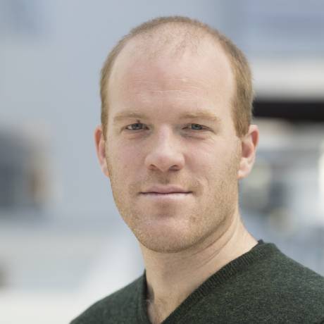
dr.ir. P. Kruizinga
Guest Assistant Professor
Signal Processing Systems (SPS), Department of Microelectronics
Signal Processing Systems (SPS), Department of Microelectronics
Expertise: Ultrasound imaging of the brain
Themes: Health and WellbeingBiography
Pieter Kruizinga is a visiting assistant professor at TU Delft, and assistant professor at Erasmus Medical Center, Department of Neuroscience.
Graph Signal Processing in Action
Graph signal processing, Graph topology identification, Graph filtering, Dynamic graphs, Graph learning, Functional ultrasound, Recommender systems
- Multiple Measurement Vector Model for Sparsity-Based Vascular Ultrasound Imaging
Dogan, D.; Kruizinga, P.; Bosch, J.G.; Leus, G.;
In Proc. of IEEE Statistical Signal Processing Workshop (SSP),
Rio de Janeiro, Brazil, pp. 501--505, July 2021. DOI: 10.1109/SSP49050.2021.9513860 - Coding Mask Design for Single Sensor Ultrasound Imaging
P. van der Meulen; P. Kruizinga; J.G. Bosch; G. Leus;
IEEE Trans. on Computational Imaging,
Volume 6, pp. 358--373, 2020. DOI: 10.1109/TCI.2019.2948729 - Blind calibration for arrays with an aberration layer in ultrasound imaging
P. van der Meulen; M. Coutino; P. Kruizinga; J.G. Bosch; G. Leus;
In 29th European Signal Processing Conference (EUSIPCO 2020),
Amsterdam (Netherlands), EURASIP, pp. 1270-1274, August 2020.
document - Joint Estimation of Hemodynamic Response and Stimulus Function in Functional Ultrasound Using Convolutive Mixtures
Aybuke Erol; Simon Van Eyndhoven; Sebastiaan Koekkoek; Pieter Kruizinga; Borbala Hunyadi;
In 2020 54th Asilomar Conference on Signals, Systems, and Computers,
IEEE, 2020. - Vector Doppler imaging of small vessels using directionally filtered Power Doppler images
B. Generowicz; L. Verhoef; F. Mastik; S. Dijkhuizen; N. van Dorp; J. Voorneveld; J. Bosch; K. Kumar; G. Leus; C. de Zeeuw; S. Koekkoek; P. Kruizinga;
In 2020 IEEE International Ultrasonics Symposium (IUS),
pp. 1-4, 2020. DOI: 10.1109/IUS46767.2020.9251356
document - A Reconfigurable Ultrasound Transceiver ASIC With 24 × 40 Elements for 3D Carotid Artery Imaging
E. Kang; Q. Ding; M. Shabanimotlagh; P. Kruizinga; Z. Y. Chang; E. Noothout; H. J. Vos; J. G. Bosch; M. D. Verweij; N. de Jong; M. A. P. Pertijs;
IEEE Journal of Solid-State Circuits,
Volume 53, Issue 7, pp. 2065-2075, July 2018. DOI: 10.1109/JSSC.2018.2820156
Abstract: ...
This paper presents an ultrasound transceiver application-specific integrated circuit (ASIC) designed for 3-D ultrasonic imaging of the carotid artery. This application calls for an array of thousands of ultrasonic transducer elements, far exceeding the number of channels of conventional imaging systems. The 3.6 x 6.8 mm² ASIC interfaces a piezo-electric transducer (PZT) array of 24 x 40 elements, directly integrated on top of the ASIC, to an imaging system using only 24 transmit and receive channels. Multiple ASICs can be tiled together to form an even bigger array. The ASIC, implemented in a 0.18 μm high-voltage (HV) BCD process, consists of a reconfigurable switch matrix and row-level receive circuits. Each element is associated with a compact bootstrapped HV transmit switch, an isolation switch for the receive circuits and programmable logic that enables a variety of imaging modes. Electrical and acoustic experiments successfully demonstrate the functionality of the ASIC. In addition, the ASIC has been successfully used in a 3-D imaging experiment. - Fast volumetric imaging using a matrix TEE probe with partitioned transmit-receive array
D. Bera; F. van den Adel; N. Radeljic-Jakic; B. Lippe; M. Soozande; M. A. P. Pertijs; M. D. Verweij; P. Kruizinga; V. Daeichin; H. J. Vos; A. F. W. van der Steen; J. G. Bosch; N. de Jong;
Ultrasound in Medicine \& Biology,
Volume 44, Issue 9, pp. 2025-2042, July 2018. DOI: 10.1016/j.ultrasmedbio.2018.05.017
Abstract: ...
We describe a 3-D multiline parallel beamforming scheme for real-time volumetric ultrasound imaging using a prototype matrix transesophageal echocardiography probe with diagonally diced elements and separated transmit and receive arrays. The elements in the smaller rectangular transmit array are directly wired to the ultrasound system. The elements of the larger square receive aperture are grouped in 4 × 4-element sub-arrays by micro-beamforming in an application-specific integrated circuit. We propose a beamforming sequence with 85 transmit–receive events that exhibits good performance for a volume sector of 60° × 60°. The beamforming is validated using Field II simulations, phantom measurements and in vivo imaging. The proposed parallel beamforming achieves volume rates up to 59 Hz and produces good-quality images by angle-weighted combination of overlapping sub-volumes. Point spread function, contrast ratio and contrast-to-noise ratio in the phantom experiment closely match those of the simulation. In vivo 3-D imaging at 22-Hz volume rate in a healthy adult pig clearly visualized the cardiac structures, including valve motion. - Structured ultrasound microscopy
J. Janjic; P. Kruizinga; P. van der Meulen; G. Springeling; F. Mastik; G. Leus; J.G. Bosch; A.F.W. van der Steen; G. van Soest;
Applied Physics Letters,
Volume 112, Issue 25, April 2018. DOI: 10.1063/1.5026863
document - Calibration techniques for single-sensor ultrasound imaging with a coding mask
P. van der Meulen; P. Kruizinga; J.G. Bosch; G. Leus;
In 52nd Asilomar Conference on Signals, Systems and Computers,
IEEE, November 2018.
document - Efficient and Flexible Spatiotemporal Clutter Filtering of High Frame Rate Images Using Subspace Tracking
B.S. Generowicz; G. Leus; S.S. Tbalvandanv; W.S. Van Hoogstraten; C. Strydis; J.G. Bosch; A.F.W. van der Steen; C.I. de Zeeuw; S.K.E. Koekkoek; P. Kruizinga;
In 2018 IEEE International Ultrasonics Symposium (IUS),
pp. 206-212, Oct. 2018. ISSN 1948-5727. DOI: 10.1109/ULTSYM.2018.8579775
document - High Frequency Functional Ultrasound in Mice
S.K.E. Koekkoek; S.S. Tbalvandany; B.S. Generowicz; W.S. van Hoogstraten; N.L. de Oude; H.J. Boele; C. Strydis; G. Leus; J.G. Bosch; A.F.W. van der Steen; C.I. de Zeeuw; P. Kruizinga;
In 2018 IEEE International Ultrasonics Symposium (IUS),
pp. 1-4, Oct. 2018. ISSN 1948-5727. DOI: 10.1109/ULTSYM.2018.8579865
document - Compressive 3D ultrasound imaging using a single sensor
P. Kruizinga; P. van der Meulen; A. Fedjajevs; F. Mastik; G. Springeling; N. de Jong; J.G. Bosch; G. Leus;
Science Advances,
Volume 3, December 2017. ISSN: 2375-2548. DOI: 10.1126/sciadv.1701423
document
Youtube - Model-based image reconstruction for medical ultrasound
P. Kruizinga; P. van der Meulen; F. Mastik; N. de Jong; J. G. Bosch; G. Leus;
The Journal of the Acoustical Society of America,
Volume 141, Issue 5, pp. 3610-3610, June 2017. DOI: 10.1121/1.4987733 - Acoustical compressive 3D imaging with a single sensor
P. Kruizinga; P. van der Meulen; F. Mastik; A. Fedjajevs; G. Springeling; N. de Jong; G. Leus; J. G. Bosch;
In 2017 IEEE International Ultrasonics Symposium (IUS),
pp. 1-1, September 2017. DOI: 10.1109/ULTSYM.2017.8091779
document - Spatial Compression in Ultrasound Imaging
P. van der Meulen; P. Kruizinga; J. G. Bosch; G. Leus;
In 51st Asilomar Conf. on Signals, Systems and Computers,
Asilomar (CA), IEEE, October 2017. - Impulse response estimation method for ultrasound arrays
P. van der Meulen; P. Kruizinga; J. G. Bosch; G. Leus;
In 2017 IEEE International Ultrasonics Symposium (IUS),
pp. 1-4, September 2017. DOI: 10.1109/ULTSYM.2017.8092977
document - A Reconfigurable 24 × 40 Element Transceiver ASIC for Compact 3D Medical Ultrasound Probes
E. Kang; Q. Ding; M. Shabanimotlagh; P. Kruizinga; Z. Y. Chang; E. Noothout; H. J. Vos; J. G. Bosch; M. D. Verweij; N. de Jong; M. A. P. Pertijs;
In Proc. European Solid-State Circuits Conference (ESSCIRC),
IEEE, pp. 211-214, September 2017. - Volumetric imaging using adult matrix TEE with separated transmit and receive array
D. Bera; F. van den Adel; N. Radeljic-Jakic; B. Lippe; M. Soozande; M. Pertijs; M. Verweij; P. Kruizinga; V. Daeichin; H. Vos; J. Bosch; N. de Jong;
In Proc. IEEE International Ultrasonics Symposium (IUS),
IEEE, pp. 1-1, September 2017. (abstract). DOI: 10.1109/ULTSYM.2017.8092906
Abstract: ...
The design of 3D TEE transducers poses severe technical challenges: channel count, electronics integration with high and low voltages, heat dissipation, etc. We present an adult matrix TEE probe with separate transmit (Tx) and receive (Rx) arrays allowing optimization in both Tx and Rx [1]. Tx elements are directly wired out, Rx employs integrated micro-beamformers in low-voltage (1.8/5.0V) chip technology. The prototype is fully integrated into a gastroscopic tube. - Towards 3D ultrasound imaging of the carotid artery using a programmable and tileable matrix array
P. Kruizinga; E. Kang; M. Shabanimotlagh; Q. Ding; E. Noothout; Z. Y. Chang; H. J. Vos; J. G. Bosch; M. D. Verweij; M. A. P. Pertijs; N. de Jong;
In Proc. IEEE International Ultrasonics Symposium (IUS),
IEEE, pp. 1-3, September 2017. DOI: 10.1109/ULTSYM.2017.8091570
Abstract: ...
Accurate assessment of carotid artery disease by measuring blood flow, plaque deformation and pulse wave velocity using ultrasound imaging requires 3D information. Additionally, the volume rates should be high enough (> 1 kHz) to capture the full range of these fast transient phenomena. For this purpose, we have built a programmable, tileable matrix array that is capable of providing 3D ultrasound imaging at such volume rates. This array contains an application-specific integrated circuit (ASIC) right beneath the acoustic piezo-stack. The ASIC enables fast programmable switching between various configurations of elements connected to the acquisition system via a number of channels far smaller than the number of transducer elements. This design also allows for expanding the footprint by tiling several of these arrays together into one large array. We explain the working principles and show the first basic imaging results of a 2-by-1 tiled array. - Optimizing the directivity of piezoelectric matrix transducer elements mounted on an ASIC
M. Shabanimotlagh; S. Raghunathan; V. Daeichin; P. Kruizinga; H. J. Vos; M. A. P. Pertijs; J. G. Bosch; N. de Jong; M. D. Verweij;
In Proc. IEEE International Ultrasonics Symposium (IUS),
IEEE, pp. 1-4, September 2017. DOI: 10.1109/ULTSYM.2017.8091752
Abstract: ...
Over the last decade, clinical studies show a strong interest in real-time 3D imaging. This calls for ultrasound probes with high-element-count 2D matrix transducer arrays. These may be interfaced to an imaging system using an in-probe Application Specific Integrated Circuit (ASIC) that takes care of signal amplification, element switching, sub-array beamforming, etc. Since the ASIC is made from silicon and is mounted directly behind the transducer elements, it can acoustically be regarded as a rigid plate that can sustain traveling lateral waves. These waves lead to acoustical cross-talk between the elements, and results in extra peaks in the directivity pattern. We propose two solutions to this problem, based on numerical simulations. One approach is to decrease the phase velocity in the silicon by reducing the silicon thickness and absorbing the energy using a proper backing material. Another solution is to disturb the waves inside the silicon plate by sub-dicing the back-side of the ASIC. We conclude that both solutions can be used to improve the directivity pattern.
BibTeX support
Last updated: 2 Oct 2023
