Medical Delta Cardiac Arrhythmia Lab (CAL)
Medical Delta Program Objectives
Cardiac arrhythmias are the cardiovascular epidemic of the 21th century. Unfortunately, effectiveness of arrhythmia therapy is low due to insufficient knowledge of underlying electropathology, which is defined as electrical conduction disorders caused by damage of cardiac tissue. Also, there are currently no tools to identify and target electropathology.
The mission of the Medical Delta team is to decrease the cardiac arrhythmia burden by providing patient-tailored therapy. Hereto, we aim to design and test novel bio-electrical diagnostic tools, unravel arrhythmia-related electropathology and develop innovative therapies targeting electropathology. This goal requires construction of bio-electrical recording and processing techniques to be utilized to unravel and target electropathology.
Therefore, we propose to
- design (multi-modal) devices for identifying arrhythmia-related electropathology, in both in-vitro cell- and tissue cultures and in-vivo animal or human hearts. This includes a first-of-a-kind arrhythmia-on-a-chip model to study arrhythmia mechanisms, identify novel therapeutic targets and test innovative therapies,
- generation of high-resolution electrical signal fingerprints of patients using advanced signal-processing techniques and
- unravelling electropathology in humans by integrating high-resolution signal fingerprints with blood- and tissue-based biomarkers and cardiac images.
In this way, we provide a worldwide unique individualized, multi-disciplinary management strategy of patients with cardiac arrhythmias.
Medical Delta 2.0 has funded two PhD projects as a start of the overall program.
Project 1 objectives: Design of (multi-modal) devices for identifying arrhythmia-related electropathology and invasive therapies
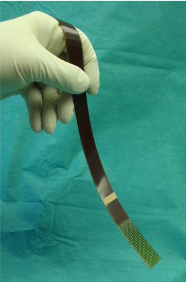
Dedicated high-resolution, flexible electrode arrays capable of both sensing and pacing with miniaturized electronics, suitable for measuring at either the inner or outer surface of the heart are at present not available. These devices are essential to unravel and target arrhythmia-related electropathology. Currently available mapping devices for both in-vitro cell and tissue structures and in-vivo animal or human hearts are limited by the size and number of sensing or pacing electrodes. Furthermore, they do not visualize underlying cardiac structure. Also, there are no devices combining arrays of ultrasound and bio-potential electrodes enabling localization of the origin of abnormal signals thereby facilitating identification of electropathology. In addition, easily applicable screening devices suitable for people of all ages for accurate, long-term detection of cardiac arrhythmias do not exist.
Challenging engineering aspects include, e.g., high-voltage stimulation techniques and precise recording electronics, multiplexing, compression/data reduction, conversion and communication techniques, artefact-reduction techniques, reliability and safety, including MRI compatibility.
The task of PhD 1 is to design and develop an innovative microelectronic system to interface with the arrhythmia-on-a-chip platform for synchronous, multi-sensing and automated measurement of electromechanical cardiac cell activity. The system will be adopted to identify electropathology-related signals in cell and tissue models of arrhythmias, particularly atrial fibrillation and to test pharmacological therapies.
Project 2 objectives: Unraveling electropathology by integrating (bio)-electrical profiles with cardiac images
To identify areas of electropathology as target sites for treatment, it is crucial to obtain detailed insights into the interrelationship between high-resolution (interelectrode distances 2mm) excitation maps, signal fingerprints, blood- and tissue-based biomarkers and clinical profiles including, e.g., echo- and MRI cardiac images. Due to its accuracy, high-resolution epicardial mapping is the foundation of the development of less (catheter-based) or non-invasive (body surface) mapping approaches to identify the arrhythmogenic substrate.
The combination of unique (non)-invasive high-resolution mapping data and cardiac images visualizing electropathology can 1) serve as a novel research tool to explore relations between signal morphologies and tissue damage, 2) serve as a novel diagnostic imaging tool to stage cardiac arrhythmias and 3) guide selection of the appropriate treatment modality, enabling patient tailored therapy.
The task of PhD 2 is to apply big data analytics to high-resolution cardiac signals obtained during normal heart rhythms and cardiac arrhythmias and to develop a strategy to combine high resolution electrical and bio-molecular data with cardiac images.
Project data
| Researchers: | Alle-Jan van der Veen, Borbála Hunyadi, Wouter Serdijn, Hanie Moghaddasi, Rui Guan, Frans Widdershoven, Natasja de Groot |
|---|---|
| Starting date: | January 2019 |
| Closing date: | July 2023 |
| Funding: | 380 kE; related to group 190 kE |
| Sponsor: | Medical Delta |
| Partners: | Erasmus MC (Prof. Dr. N.M.S. de Groot, Prof. Dr. B. Brundel) |
| Contact: | Borbála Hunyadi |