Novel multimodal endoscopic probes for simultaneous PET/ultrasound imaging for image-guided interventions (EndoTOFPET-US)
Moreover, significant gain in sensitivity is to be expected from improvements in timing performance and detector miniaturization (allowing imaging closer to the target). Such gains will lead to detection and precise localisation of smaller tumours, enabling faster exams with higher patient throughput, decreased image degradation from moving organs and significant radiation dose reduction. In particular such advantages will play a major role in the earlier detection and patient-tailored treatment of rather asymptomatic cancer types improving potentially their prognosis.
In a parallel dimension, these novel tools will allow highly specific biomarkers to be developed by improved imaging capabilities over the state-of-the-art, resulting thus in faster, more successful and more efficient in vivo evaluation.
The objectives of the Endo-TOFPET-US project are briefly described below:
- Build a detection head behaving as an optical photon counter and providing a quantum detection scheme for photons over a wide spectral range (from visible to gamma -rays) and a fully digital signal at the level of the photo-detector.
- Reach a 200ps timing resolution, corresponding to a spatial resolution of 3 cm along the Line of Response (LOR) in a dual-head system, allowing a direct (and therefore fast) 3-dimensional reconstruction through a limited number of angular projections, and an efficient rejection of background coming from outside of the few cubic centimetres region of interest (ROI).
- Achieve a millimetre spatial resolution for the PET detector head.
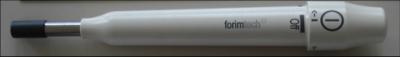
- Integrate all the components in a very compact detector head with an efficient tracking system for a sub-millimetre on-line determination of the position of the different imaging parts.
- Generate fully 3D tomographic images and fuse them with the US images for hybrid visualization of the biomarker distribution (PET) and the anatomy (US).
- Define a roadmap for the development of a new generation of multimodal endoscopic probes combining the morphological information with submillimeter spatial resolution provided by the US endoscopic probe with the high-sensitivity measurement of the functional response of specific radioactive biomarkers acquired by a dedicated high performance and versatile PET camera.
For more information, see the
project homepage.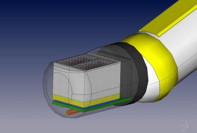
Project data
| Researchers: | Edoardo Charbon, Shingo Mandai, Tim Gong |
|---|---|
| Starting date: | January 2011 |
| Closing date: | July 2015 |
| Sponsor: | EU FP7 |
| Partners: | Aix-Marseille University (France), Centre Hospitalier Universitaire Vaudois et Universite de Lausanne (Switzerland), CERN (Switzerland), LIP (Portugal), Fibercryst, Kloe, SurgicEye, TU Munchen, University of Heidelberg, University Milano Biccoca |
| Contact: | Edoardo Charbon |