MSc thesis project proposal
[2020; only 1 spot left] CMOS circuit design for ultrasound based nerve activation/inhibition
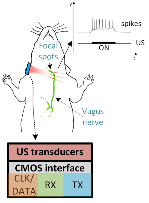
Historically, electrical stimulation has been the preferred method to interface with the central (CNS) and peripheral nervous system (PNS), and in the case of the Vagus nerve, is typically implemented by implanting a stimulator near the neck where the nerve is most accessible. Even though this therapy has been proven effective for a great number of diseases (rheumatoid arthritis, Crohn’s disease, epilepsy, depression), there are still several limitations that can further be improved. Electrical stimulation does not currently have high enough spatial resolution to target only desired bundles of nerve fibers (as small as a few µm) and, therefore, unwanted vagus nerve fibers are also affected by the stimulation, which can lead to undesired responses manifested as several side effects, such as shortness of breath, cough, hoarseness and heart-rate variations. In addition, and perhaps more importantly, this therapeutic modality requires surgery, which due to all the risks associated with it, makes this type of implantable devices still used as a last resort therapy, when pharmaceuticals stop having a positive effect.A method to achieve superior stimulation spatial resolution while remaining non-invasive is with ultrasound neuromodulation techniques [1,2]. Focused ultrasound has been shown to be effective in stimulation of both the CNS, and PNS, including the vagus nerve. This method takes advantage of the deep penetration of ultrasound energy in soft body tissue, combined with mm to sub-mm wavelengths and very low scattering, allowing for precise generation of ultrasound focal spots where nerve activation of inhibition occurs. However, existing works use commercially available single-element focused ultrasound transducers, which are bulky, require rack-mounted off-the-shelf equipment and do not allow focal spot beam steering or ultrasound imaging.
This project will focus on implementing a CMOS circuit interface for a 2D array of micron-scale ultrasound transducers. This device has the potential to enable a drastic improvement of existing ultrasound neuromodulation methods, by enabling electronic beamforming to control the location of the focal spot by software, and to allow for concurrent structural imaging of the target nerve. In the limit, this device would be able to allow simultaneous activation or inhibition of different nerve bundles, by moving the focal spot location at a much higher rate than the time constant of neuronal responses, all of this non-invasively in a wearable patch.
An existing prototype for this application has been developed and tested [3], featuring CMOS circuit with a 2D array of beamforming transmitter circuits operating at 8.5 MHz, allowing focal spot steering in 3 dimensions, with a focal spot size of ~ 200 µm x 200 µm x 600 µm. This project would enable the improvement of existing work by increasing the frequency to 25 MHz (1/3 of the wavelength leading to 3x better spatial resolution), and also including ultrasound imaging circuitry, to allow for concurrent neuromodulation and structural imaging, for focal spot targeting and nerve motion tracking.
This project can welcome up to three students, which would implement the three main building blocks of the chip: transmit beamforming circuitry, receiver circuitry for imaging, and clock/data management circuitry.
References:
[1] Yusuf Tufail, Alexei Matyushov, Nathan Baldwin, Monica L. Tauchmann, Joseph Georges, Anna Yoshihiro Stephen I. Helms and Tillery William J. Tyler, “Transcranial pulsed ultrasound stimulates intact brain circuits.”, Neuron 66 (5), 2010.
[2] Matthew E Downs, Stephen A Lee, Georgiana Yang, Seaok Kim, Qi Wang and Elisa E Konofagou, “Non-invasive peripheral nerve stimulation via focused ultrasound in vivo”, Physics in Medicine and Biology, 66 (3), 2018.
[3] Tiago Costa, Chen Shi, Kevin Tien, and K. L. Shepard, “A CMOS 2D transmit beamformer with integrated PZT ultrasound transducers for neuromodulation,” Custom Integrated Circuits Conference, 2019
Assignment
1st part: Literature review of both ultrasound neuromodulation and ultrasound phased array circuit interfaces.
- TX circuits: What pressure is required to achieve neuromodulation? What types of transducers can I use to achieve such pressures? What is its electrical model? What circuit topologies are most suited to interface with such model? How to implement a transmit beamforming circuit?
- RX circuits: What signal-to-noise ratio can I obtain when imaging the vagus nerve with ultrasound? What type of pre-amplifier is most suited? What signal processing functions do need to be implemented?
- CLK/Data management: how many transmit/receiver channels do I need to successfully interface with the vagus nerve? How many clock domains is required for both TX and RX blocks? How to digitally control the transmit beamformers?
2nd part: CMOS Circuit design implementation of the different system blocks:
- TX Circuits: analog and mixed-signal circuit design: Delay locked loops, phase rotators, level shifters, power amplifiers.
- RX Circuits: mostly analog circuit design: voltage/charge pre-amplifiers, variable-gain amplifiers, band-pass filters, analog-to-digital converters.
- CLK/DATA management: Digital circuit design: digital state machines, memories, serial interfaces.
Requirements
MSc EE-ME studentContact
dr. Tiago Costa
Bioelectronics Group
Department of Microelectronics
Last modified: 2021-02-10